
Some years ago, I broke my elbow, and a radiography of my aching limb was taken to reveal the small inner details of my bones, with my slightest discomfort. This marvel was possible because, in 1895 studying the effect of an electric discharge through a gas at low pressure, Willhelm Roentgen accidentally discovered a new kind of radiation, which he called X-rays. He correctly discovered the mechanisms of the generation of the X-rays and produced the very first radiography placing an object, not quite a random one but the hand of his wife Bertha!, between the X-ray source and a film covered with barium platinocyanide. Not much time passed before X-rays were used in medicine, in the treatment of injuries of soldiers on the WWI front, because they helped the surgeons in the localization of the bullets or shrapnel.
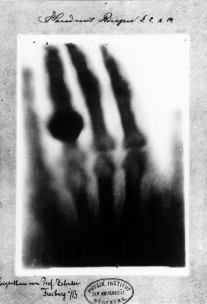
First medical X-ray by Wilhelm Röntgen of his wife Anna Bertha Ludwig’s hand
Going back to my elbow, unfortunately it was imaged in the wrong position, or in a position where my discomfort was yes minimal, but all the bones were superimposed, and the radiologist could not understand whether it was broken or not. (It was in fact broken as the doctor soon realized after I screamed with pain in her ear as she manipulated it.) Radiography, in fact gives a 2-dimensional (2D) representation of a 3D object, effectively “flattening” it, losing the depth information. Wouldn’t it be great to retain such depth information? Well, it turns out that we can! with a technique, which we can consider an “extension” of radiography, called tomography –from ancient Greek writing (graphos) slices (tomos). To be able to obtain the 3D representation of the object, one needs to gather data from more points of view; a bit like having 2 eyes –2 points of view– allows us to get depth view. This is done by “simply” rotating the object under investigation with respect to the X-ray source and detector assembly and acquiring many radiographies at different rotation angles.
The detector is like the camera in your phone, but it is sensitive to X-rays rather than visible light. In material science and engineering it is sufficient to rotate the sample, while in medical imaging, since rotating patients is not an option, it is the source-detector assembly to rotate around them. So, being subject to a tomography scan involves receiving much more X-ray radiation than for a single radiography, and that is why the latter is still extremely popular in medicine.
However, it took some 80 years of developments in mathematics, detector technology and computers to get from Roentgen’s radiographies to the beginning of the Computed Tomography (CT) era. In 1917 Johann Radon demonstrated that it is possible to obtain a 3D representation of an object from a stack of radiographies. And in the 70s, here in the UK, Godfrey Hounsfield developed the very first computer-aided tomography machine at EMI, the record company! In today’s society, tomography is used in a wide range of fields, from basic research to medical imaging, from manufacturing to security.
With my team, I write software for the processing and analysis of CT data, giving support to 2 UK academic networks related to tomography: Collaborative Computational Project (CCP) in Tomographic Imaging (CCPi) and CCP in Synergistic Image Reconstruction for Biomedical Imaging (CCP SyneRBI).The former is for material science while the latter is for medical imaging applications. The Open Source Software (OSS) we develop, the Core Imaging Library (CIL) and the Synergistic Image Reconstruction Framework (SIRF), are simple enough to be used for training and teaching, but also powerful enough to be used in research with real data.
The key processing step that brings us from the stack of radiographies to the volumetric representation is called reconstruction, and our software CIL and SIRF do just that for X-Ray CT and PET/MRI respectively. Now, I have not talked about Positron Emission Tomography (PET), nor (nuclear) Magnetic Resonance Imaging (MRI) as it would make this post too long, but these are two techniques using different mechanisms to obtain the volumetric information: gamma rays from a positron annihilation and the resonance of the proton of water excited by a microwave in a strong magnetic field, very exciting!
Several techniques exist for reconstruction: the most widely used is an analytic method named Filtered Back Projection (FBP) which is fast and very reliable. It has the drawback of requiring a number of radiographies proportional to the number of pixels in the detector and, that it does not perform well when the signal-to-noise ratio is low. CIL gives the user access to a wealth of tools to perform the reconstruction, that span from readers to import the data from an X-ray CT scanner, to algorithms to reconstruct and visualise the data. CIL also allows the creation of bespoke iterative reconstruction algorithms thanks to its modular design. Some more in-depth posts will probably follow on the subject, but if you are interested here are 2 articles describing CIL [1], [2].
Without computers and software, tomography would not exist, but increasingly all branches of science and society are reliant on the availability of digital computers and of software running on them, from supercomputers to ATMs, from mobile phones to defibrillators. We put a lot of trust in software (if not our own lives), implicitly believing that the output of such software is both correct and consistent, i.e. it is reproducible. The Software Sustainability Institute, writing about scientific software, words which could be extended to any software, says “The reproducibility of research is at the very heart of the scientific method. As more research is based on results that are generated by software, there must be an increased focus on developing software that is reliable and which can be easily proven to produce reproducible results”.[3]
CIL and SIRF are both Open Source Software, because we (the CCPi and CCP-SyneRBI communities) believe that OSS can guarantee reproducibility, being easily available, shared and especially reviewed. I suggest you to have a look at this interesting talk by Karen Sandler at the Open Source Convention 2011 because it’s not just scientific software that should be reproducible!
Let me conclude with a little note: scientific research often opens paths that are unimaginable and that is why it is important to fund basic research even if it looks like it does not give benefits outside its field. A minimum of 4 Nobel prizes have been awarded for the techniques that are cited in this post, demonstrating the high impact and importance of the basic research that was carried out.
For the discovery of the X-Rays, Roentgen was awarded the very first Nobel Prize in Physics in 1901 [4], and in 1905 Philipp E. A. von Lenard, whose device Roentgen was using while making his discovery, was awarded a Nobel Prize in Physics for his work on cathode rays [5].
In 1979, Godfrey Hounsfield and Allan Cormack were awarded the Nobel Prize in Physiology or Medicine for the development of computer assisted tomography [6].
The Nobel Prize in Physiology or Medicine 2003 was awarded to Paul Lauterbur and Sir Peter Mansfield for their discoveries concerning magnetic resonance imaging [7].
And finally, if you want to reproduce it, the code to make the chocolate egg reconstruction can be found here.
[1] Jørgensen JS et al. 2021 Core Imaging Library Part I: a versatile python framework for tomographic imaging. Phil. Trans. R. Soc. A 20200192.
Code at https://github.com/TomographicImaging/Paper-2021-RSTA-CIL-Part-I
[2] Papoutsellis E et al. 2021 Core Imaging Library – Part II: multichannel reconstruction for dynamic and spectral tomography. Phil. Trans. R. Soc. A 20200193.
Code at https://github.com/TomographicImaging/Paper-2021-RSTA-CIL-Part-II
[3] https://www.software.ac.uk/about/manifesto
[4] https://www.nobelprize.org/prizes/physics/1901/summary/
[5] https://www.nobelprize.org/prizes/physics/1905/summary/
[6] https://www.nobelprize.org/prizes/medicine/1979/summary/
[7] https://www.nobelprize.org/prizes/medicine/2003/summary/